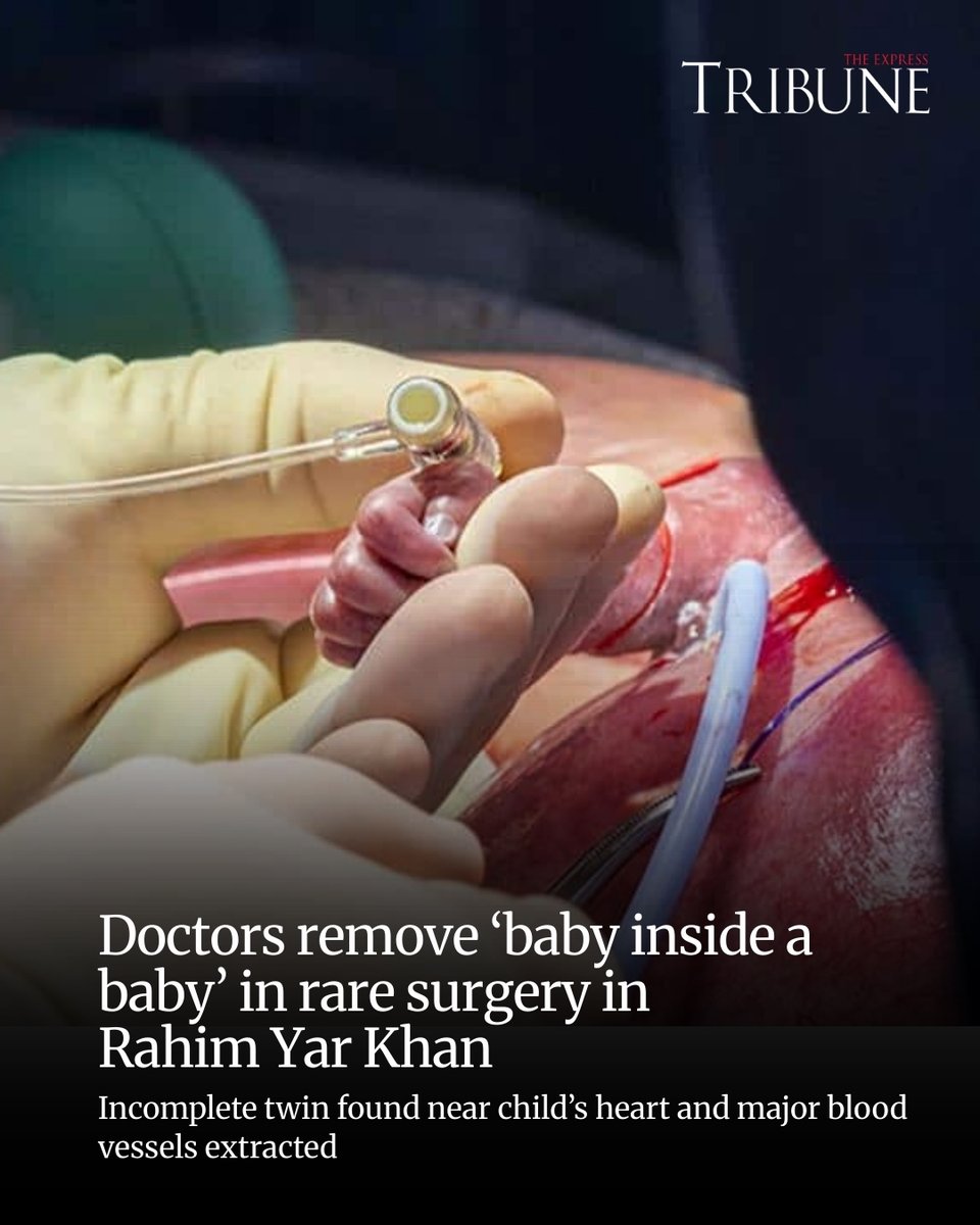
Fetus-in-Fetu Surgery: Rare Anomaly Removed from Boy’s Chest
In a truly extraordinary medical achievement, doctors at Sheikh Zayed Medical College and Hospital successfully performed Fetus-in-Fetu surgery. They removed a rare ‘fetus in fetu’ from a five-year-old boy’s chest cavity. This uncommon congenital anomaly presented a significant challenge. The underdeveloped twin was lodged dangerously close to vital organs. Consequently, this successful surgery became a testament to remarkable medical precision and expertise.
Young Rehan, the patient, arrived at the hospital suffering from a persistent cough. He also experienced increasingly severe breathing difficulties. Initial diagnostic scans unveiled a shocking truth: a partially developed fetus resided within his chest. This condition occurs in only about one in 500,000 births worldwide. Furthermore, chest presentations are exceptionally rare, making this case truly unique.
Fetus-in-Fetu: Understanding this Rare Medical Enigma
Fetus-in-fetu is a fascinating and complex condition. It involves a malformed parasitic twin found within the body of its sibling. Experts believe it originates during early embryonic development. Often, an identical twin fails to separate completely. Instead, it becomes enveloped by the developing host twin. The parasitic twin often contains recognizable structures like limbs, bones, or even organs. However, it is typically anencephalic (lacking a brain) and achardiac (lacking a heart), rendering it non-viable.
Most documented cases of fetus-in-fetu are discovered in the abdominal cavity. However, its occurrence within the thoracic region, or chest, is exceedingly rare. This particular location adds layers of complexity and risk. The parasitic mass can compress and interfere with critical organs. For example, it can affect the heart and lungs, posing a direct threat to the host child’s life.

Pre-Surgical Challenges and Expert Team Collaboration
Diagnosing fetus-in-fetu, especially in such an unusual location, required meticulous assessment. Rehan’s parents had consulted numerous doctors previously. Nevertheless, the true nature of his ailment remained elusive until advanced imaging at Sheikh Zayed Medical College. The fetus’s presence near the heart and major blood vessels meant intervention would be perilous. Therefore, a highly skilled and experienced surgical team was absolutely essential.
Senior Thoracic Surgeon Dr. Sultan Mahmood spearheaded this complex surgical procedure. His leadership and the collective expertise of the medical team proved crucial. They meticulously navigated the delicate anatomy of a five-year-old’s chest cavity. A single misstep could have led to catastrophic consequences. Consequently, their precision ensured a successful outcome.
High-Stakes Operation: A Triumph of Surgical Precision
The operation to remove the malformed fetus was a high-stakes endeavor. It demanded extreme precision and meticulous planning. Dr. Sultan Mahmood emphasized the delicate nature of the Fetus-in-Fetu surgery. He highlighted the fetus’s proximity to vital structures. This necessitated extraordinary care to avoid damage to the heart, major arteries, or lungs. The surgical team worked for several hours. They carefully dissected and extracted the mass. Meanwhile, they ensured Rehan’s stability throughout the entire procedure.
The successful removal of the fetus alleviated Rehan’s immediate health risks. Furthermore, it prevented potential long-term complications associated with its continued presence. This surgical triumph highlights advancements in medical technology. It also underscores the profound impact of dedicated healthcare professionals.

Rehan’s Promising Recovery after Fetus-in-Fetu Surgery
Following the successful Fetus-in-Fetu surgery, Rehan is now on a promising path to recovery. He receives advanced care in the thoracic ward. His mother expressed immense gratitude to the medical team. She praised their dedication and skill in saving her son’s life. This followed a long and confusing journey for their family. Hospital spokesperson Dr. Ilyas Rana confirmed Rehan’s stable condition and ongoing recovery. This underscores the significance of this particular case.
This specific case of fetus-in-fetu in the chest is exceptionally rare globally. Its successful resolution represents a significant contribution to medical literature. Moreover, it offers hope for families facing similar daunting diagnoses. It serves as a powerful reminder of continuous strides made in surgical capabilities and diagnostic technologies.

A Beacon of Hope in Rare Medical Conditions
The successful removal of the fetus-in-fetu from Rehan’s chest stands as a remarkable achievement. It is a credit to Sheikh Zayed Medical College and Hospital and the medical community at large. This case showcases the critical importance of early diagnosis and advanced imaging techniques. Furthermore, it highlights the unparalleled skill of surgical teams. They are capable of tackling conditions once considered insurmountable.
Rehan’s story is a beacon of hope. It illustrates how perseverance and modern medicine can overcome even the rarest and most challenging health conditions. It underscores the profound impact dedicated medical professionals have on individuals and their families. This ensures that even amidst unique medical mysteries, a healthy future is possible.